Pulmonary Embolism
What is a pulmonary embolism?
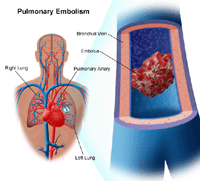
A pulmonary embolism (PE) is a blood clot that develops in a blood vessel elsewhere in the body (most commonly from the leg), travels to an artery in the lung, and forms an occlusion (blockage) of the artery.
A blood clot (thrombus) that forms in a blood vessel in one area of the body, breaks off, and travels to another area of the body through the bloodstream is called an embolus. An embolus can lodge itself in a blood vessel, blocking the blood supply to a particular organ. This blockage of a blood vessel by an embolus is called an embolism.
An embolism to the lung may cause serious life-threatening consequences and, potentially, death. Most commonly, a PE is the result of a condition called deep vein thrombosis (blood clot in the deep veins of the leg).
 |
| Click Image to Enlarge |
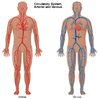
The circulatory system
The heart, arteries, capillaries, and veins make up the body's circulatory system. Blood is pumped with great force from the heart into the arteries, then into the capillaries (small blood vessels in the tissues) and returns to the heart through the veins. Much of the force of the heartbeat is lost when the blood enters the veins and results in the slowing down of the blood flow through the veins back to the heart. Under certain conditions, decreased blood flow may contribute to clot formation.
What causes a pulmonary embolism?
Blood clotting is a normal process that occurs in the body to prevent bleeding. The body makes blood clots and then breaks them down. Under certain circumstances, the body may be unable to break down a clot, which may result in a serious health condition.
Abnormal blood clotting in the veins is related to a combination of several problems such as "sluggish" blood flow through the veins, an abnormality in clot forming factors, and/or an injury to the blood vessel wall.
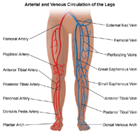
Blood clots can form in arteries and/or veins. Clots formed in veins are called venous clots. Veins of the legs can be classified as superficial veins (close to the surface of the skin) or deep veins (located near the bone and surrounded by muscle).
 |
| Click Image to Enlarge |
Venous clots most often occur in the deep veins of the legs. This condition is called deep vein thrombosis (DVT), or deep vein clot. Once a clot has formed in the deep veins of the leg, there is a potential for part of the clot to break off and travel (embolize) through the bloodstream to another area of the body. Deep vein thrombosis is the most common cause of a pulmonary embolism. Therefore, the term venous thromboembolism (VTE) may refer to deep vein thrombosis and/or the complication, pulmonary embolism.
Other less frequent sources of pulmonary embolism are a fat embolus, amniotic fluid embolus, air bubbles, and a deep vein thrombosis in the upper body. Clots may also form on the end of an indwelling intravenous (IV) catheter, break off, and travel to the lungs.
What are the risk factors for pulmonary embolism?
Risk factors that are associated with the processes that may increase the risk of a venous thromboembolism include:
-
Genetic conditions that increase the risk of blood clot formation
-
Surgery or trauma (especially to the legs) or orthopedic surgery
-
Situations in which mobility is limited, such as extended bed rest, flying or riding long distances, or paralysis
-
Previous history of clots
-
Older age
-
Cancer and cancer therapy
-
Certain medical conditions, such as heart failure, chronic obstructive pulmonary disease (COPD), hypertension (high blood pressure), stroke, and inflammatory bowel disease (chronic inflammation of the digestive tract)
-
Certain medications, such as hormone replacement therapy (estrogen pills for postmenopausal women)
-
Pregnancy (during and after pregnancy, including cesarean section)
-
Obesity
-
Varicose veins (enlarged veins in the legs)
-
Cigarette smoking
A risk factor is anything that may increase a person's chance of developing a disease. It may be an activity, such as smoking, diet, family history, or many other things. Different diseases have different risk factors.
Although these risk factors increase a person's risk, they do not necessarily cause the disease. Some people with one or more risk factor never develop the disease, while others develop disease and have no known risk factors. Knowing your risk factors to any disease can help to guide you into the appropriate actions, including changing behaviors and being clinically monitored for the disease.
What are the symptoms of a pulmonary embolism?
The following are the most common symptoms for pulmonary embolism. However, each individual may experience symptoms differently:
-
Sudden shortness of breath (most common)
-
Chest pain (usually worse with breathing)
-
A feeling of anxiety
-
A feeling of dizziness, lightheadedness, or fainting
-
Palpitations (heart racing)
-
Coughing up blood (hemoptysis)
-
Sweating
-
Low blood pressure
-
Symptoms of deep vein thrombosis, such as:
-
Pain in the affected leg (may occur only when standing or walking)
-
Swelling in the leg
-
Soreness, tenderness, redness, and/or warmth in the leg(s)
-
Redness and/or discolored skin
You may or may not have these symptoms should a pulmonary embolism occur. Usually, if a PE is suspected, the physician will check your legs for evidence of a deep vein thrombosis.
The type and extent of symptoms of a pulmonary embolism will depend on the size of the embolism and whether the person already has existing heart and/or lung problems.
The symptoms of a pulmonary embolism may resemble other medical conditions or problems. Always consult your doctor for a diagnosis.
How is pulmonary embolism diagnosed?
 |
| Click Image to Enlarge |
Pulmonary embolism is often difficult to diagnose because the signs and symptoms of PE mimic those of many other conditions and diseases.
In addition to a complete medical history and physical examination, diagnostic procedures for a pulmonary embolism may include any, or a combination, of the following:
-
Chest X-ray. A type of diagnostic radiology procedure used to assess the lungs, as well as the heart. Chest X-rays may provide important information regarding the size, shape, contour, and anatomic location of the heart, lungs, bronchi, great vessels (aorta and pulmonary arteries), and mediastinum (area in the middle of the chest separating the lungs).
-
Ventilation-perfusion scan (V/Q scan). A type of nuclear radiology procedure in which a tiny amount of a radioactive substance is used during the procedure to assist in the examination of the lungs. A ventilation scan evaluates ventilation, or the movement of air into and out of the bronchi and bronchioles. A perfusion scan evaluates blood flow within the lungs.
-
Pulmonary angiogram. An X-ray image of the blood vessels used to evaluate various conditions, such as aneurysm, stenosis (narrowing of the blood vessel), or blockages. A dye (contrast) will be injected through a thin flexible tube placed in an artery. This dye makes the blood vessels visible on X-ray.
-
Spiral computed tomography (also called CT or CAT scan). A diagnostic procedure that uses a combination of X-rays and computer technology to produce horizontal, or axial, images (often called slices) of the body. CT with contrast enhances the image of the blood vessels in the lungs. Contrast refers to a substance injected into an intravenous (IV) line that causes the particular organ or tissue under study to be seen more clearly.
-
Magnetic resonance imaging (MRI). A diagnostic procedure that uses a combination of large magnets, radiofrequencies, and a computer to produce detailed images of organs and structures within the body.
-
Duplex ultrasound (US). A type of vascular ultrasound procedure done to assess blood flow and the structure of the blood vessels in the legs. Blood clots from the legs often dislodge into the lung. Since the treatment of DVT or deep venous thrombosis and PE are the same, US is a portable, less risky and cheaper alternative that gives your doctor the same information.
-
Laboratory tests. Blood tests to check the blood's clotting status, including a test called D-dimer level. Other blood work may include testing for genetic (inherited) disorders that may contribute to abnormal clotting of the blood. In addition, arterial blood gases may be checked to determine the amount of oxygen in the blood.
-
Electrocardiogram (ECG or EKG). One of the simplest and fastest procedures used to evaluate the heart. Electrodes (small, plastic patches) are placed at certain locations on the chest, arms, and legs. When the electrodes are connected to an ECG machine by lead wires, the electrical activity of the heart is measured, interpreted, and printed out for the doctor's information and further interpretation.
Treatment for pulmonary embolism
Specific treatment will be determined by your doctor based on:
-
Your age, overall health, and medical history
-
Extent of the disease
-
Your signs and symptoms
-
Your tolerance for specific medications, procedures, or therapies
-
Expectations for the course of the disease
-
Your opinion or preference
Treatment options for pulmonary embolism include:
-
Anticoagulants. Also described as blood thinners, these medications decrease the ability of the blood to clot. Examples of anticoagulants include warfarin (Coumadin) and heparin.
-
Fibrinolytic therapy. Also called clot busters, these medications are given intravenously (IV) to break down the clot.
-
Vena cava filter. A small metal device placed in the vena cava (the large blood vessel that returns blood from the body to the heart) may be used to prevent clots from traveling to the lung. These filters are generally used in patients who cannot receive anticoagulation treatment (for medical reasons), who develop additional clots even with anticoagulation treatment, or who develop bleeding complications from anticoagulation.
-
Pulmonary embolectomy. Surgical removal of a pulmonary embolism. This procedure is generally performed only in severe situations in which the PE is very large, the patient either cannot receive anticoagulation and/or thrombolytic therapy due to other medical considerations or has not responded adequately to those treatments, and the patient's condition is unstable.
-
Percutaneous thrombectomy. Insertion of a catheter (long, thin, hollow tube) to the site of the embolism, using X-ray guidance. Once the catheter is in place, the catheter is used to break up the embolism, extract it (pull it out), or dissolve it by injecting thrombolytic medication.
An important aspect of treatment of pulmonary embolism is prophylactic (preventive) treatment to prevent formation of additional embolisms.
Prevention of pulmonary embolism
Because pulmonary embolism is caused by an embolus formed elsewhere in the body (generally in the legs), and because it is often difficult to detect presence of a venous embolus prior to the onset of complications, such as a pulmonary embolism, the prevention of these emboli is necessary in the prevention of PE.
In order to prevent pulmonary embolism, the only effective way is to prevent deep vein thrombosis. Prophylactic treatment to prevent DVT includes:
Many patients remain at risk for development of DVT for a period of time after they are either discharged from the hospital or transferred to a different type of care facility. It is important that prophylactic treatment for DVT continue until the risk for DVT development has been resolved.